Chapter 5: Skin Appendages: Hair, Glands & Nails
The appendages (adneksa) of the skin are structures that grow down from the epidermis into the dermis during development: hair follicles, sebaceous glands, sweat glands and nails. They are epidermal in origin, so even deep in the dermis they are lined by epithelium, and they are the source of new epidermis when the surface is destroyed (for example after a burn that spares the deep dermis).
Hair

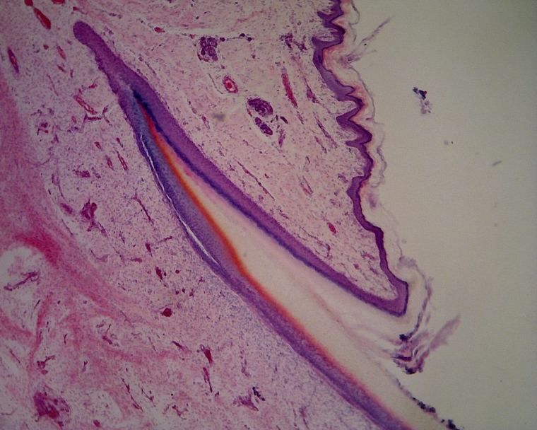
A hair is divided into:
- the hair shaft, the part above the skin surface;
- the hair root, the part within the skin, enclosed by the hair follicle;
- the hair bulb, the expanded base of the follicle, containing:
- the dermal papilla, a knob of connective tissue from the dermis that pushes up into the bulb;
- numerous blood vessels, which feed the growing hair;
- epithelial cells (the hair matrix), the site of regeneration, which divide to produce the hair;
- melanocytes among the epithelial cells, which give the hair its color.
Layers of the hair itself
The hair shaft consists of three concentric layers, from the inside out:
- Medulla: a core of loosely arranged, partly keratinized cells (absent in fine hairs).
- Cortex: the bulk of the hair, hard keratinized cells with most of the pigment.
- Cuticle: a single layer of flat, overlapping keratinized cells like roof tiles.

The follicle's sheaths
In general, the sheath around the hair root consists of two main parts, separated by the glassy membrane (a thick basement membrane):
- Dermal root sheath (connective tissue sheath): the outer layer, made of connective tissue similar to the dermis. It is the attachment site for one end of the arrector pili muscle.
- Epidermal root sheath: the inner layer, made of epithelium similar to the epidermis. It is subdivided into an outer root sheath (continuous with the epidermis) and an inner root sheath (made of Henle's layer, Huxley's layer and the sheath cuticle, next to the hair).

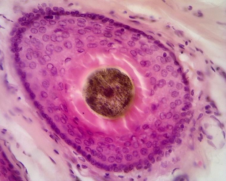
Arrector pili
The arrector pili is a small bundle of smooth muscle running from the dermal root sheath to the papillary dermis. When it contracts (sympathetic stimulation: cold or fear), it pulls the hair upright and dimples the skin ("goosebumps"), and it helps squeeze sebum out of the sebaceous gland that lies in the angle between the muscle and the follicle.
Glands of the skin
Skin has two families of glands: sebaceous glands, which make an oily secretion, and sweat glands, which come in two kinds, eccrine and apocrine. The way each releases its product (merocrine, apocrine or holocrine secretion) was introduced in Epithelial Junctions, Surface Specializations & Glands in the Block 1.1 histology ebook.
Sebaceous glands
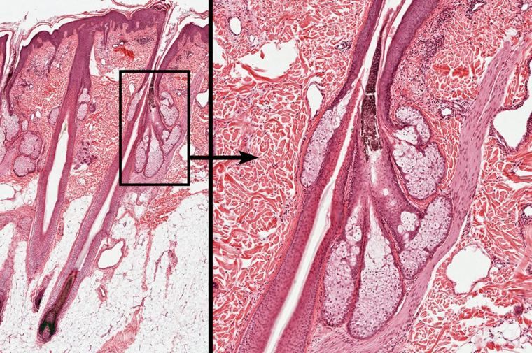
- Simple branched acinar glands.
- They open into the hair follicle; the follicle, its sebaceous gland and its arrector pili together form the pilosebaceous unit.
- Holocrine secretion: the whole cell fills with lipid, dies and disintegrates, and becomes the secretion (sebum). New cells are produced by a layer of small basal cells at the edge of each acinus.
- The secretory cells look large and pale, with a foamy cytoplasm, becoming more disrupted toward the center of the acinus.
- Present everywhere there is hair, and absent from thick skin (palms and soles). Sebum oils the hair and skin; overactivity and blockage are the basis of acne.
Eccrine sweat glands
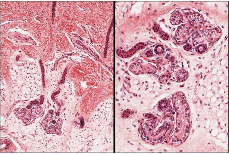
- Simple coiled tubular glands, found in almost all skin and most numerous on the palms, soles and forehead.
- The secretory portion (pars sekretorius) lies coiled in the deep dermis; a duct (pars ekskretorius) runs up through the dermis and spirals through the epidermis to open at a sweat pore on the skin surface.
- Merocrine secretion: the product leaves by exocytosis, and the cell stays intact.
- The secretory portion contains two kinds of secretory cells, dark cells (mucoid) and clear cells (which produce the watery part of sweat), plus myoepithelial cells.
- The duct has two layers of cuboidal cells (stratified cuboidal epithelium) with a narrow lumen and stains darker than the secretory part.
- Sweat cools the body by evaporation; eccrine glands are the key organs of thermoregulation.
Myoepithelial cells
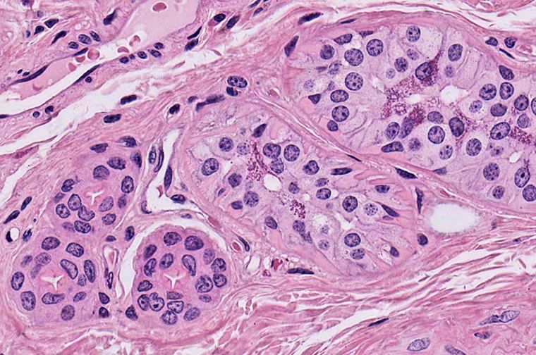
Myoepithelial cells are flattened, contractile epithelial cells that wrap around the secretory portion of sweat glands (and of other glands such as the salivary and mammary glands). They lie between the secretory cells and the basal lamina, and their contraction squeezes the secretion into the duct. They are found in the secretory portion, not in the duct.
Apocrine sweat glands
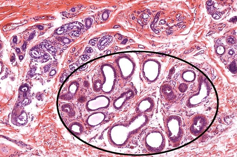
- Also coiled tubular glands, but larger, with a much wider lumen.
- They open into hair follicles (above the sebaceous gland), not onto the skin surface.
- Found only in certain regions: the axilla, the areola of the breast and the anogenital region.
- Their secretion is viscous and odorless until skin bacteria break it down, producing body odor. They become active at puberty.
- Despite the name, they are now known to secrete mainly by the merocrine route. Like eccrine glands, their secretory portion has myoepithelial cells.
| Sebaceous | Eccrine sweat | Apocrine sweat | |
|---|---|---|---|
| Shape | Simple branched acinar | Simple coiled tubular | Coiled tubular, wide lumen |
| Opens into | Hair follicle | Skin surface (sweat pore) | Hair follicle |
| Secretion | Holocrine (sebum) | Merocrine (watery sweat) | Viscous (mainly merocrine) |
| Where | Hairy skin; not palms/soles | Almost everywhere | Axilla, areola, anogenital |
| Recognize by | Large pale foamy cells | Small tubules, pale secretory + dark ducts | Large tubules, wide lumens |
Nails
The nail is a plate of hard keratin on the dorsal surface of the distal phalanx:
- The nail plate rests on the nail bed (epidermis without a granular or cornified layer, over dermis).
- Its proximal end, the nail root, lies under a fold of skin (the proximal nail fold). The nail matrix beneath the root is where the plate is produced; the pale lunula is its visible edge.
- The eponychium (cuticle) is the layer of stratum corneum extending from the proximal nail fold onto the plate; the hyponychium is the thickened epidermis under the free distal edge.
- Nails grow about 3 mm a month (fingernails), faster than toenails.