Chapter 4: Dermis, Hypodermis & Sensory Endings
Below the epidermis lies the dermis, the connective tissue that gives skin its strength and elasticity, carries its blood vessels and nerves, and houses the roots of its appendages. Below that is the hypodermis, a cushion of fat that joins the skin to the structures underneath. This chapter covers both, and then the sensory nerve endings scattered through them: the reason the skin is the body's largest sense organ.
The dermis
The dermis has two layers that differ in thickness and in the kind of connective tissue they are made of.
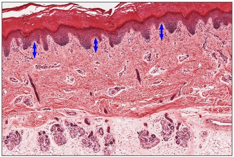
Stratum papillare (papillary layer)
- A thinner layer.
- Made of loose connective tissue.
- Contains:
- fibroblasts, macrophages and mast cells;
- the dermal papillae, which interdigitate with the epidermal ridges (Chapter 3);
- blood vessels (capillary loops) that nourish the avascular epidermis by diffusion;
- anchoring fibrils of type VII collagen, which tie the epidermal basal lamina to the dermis;
- Meissner's corpuscles (touch receptors) in the papillae.
Stratum reticulare (reticular layer)
- A much thicker layer, making up most of the dermis.
- Made of dense irregular connective tissue, mainly:
- collagen fibers (type I) in thick, interwoven bundles, which give the skin its tensile strength;
- elastic fibers, which let it recoil after stretching.
- Hair follicles, sweat and sebaceous glands, and larger vessels and nerves lie here.
| Papillary layer | Reticular layer | |
|---|---|---|
| Thickness | Thin | Thick (most of the dermis) |
| Connective tissue | Loose | Dense irregular |
| Fibers | Fine collagen (types I and III), type VII anchoring fibrils | Coarse type I collagen, elastic fibers |
| Contents | Capillary loops, Meissner's corpuscles, immune cells | Appendages, larger vessels, Ruffini endings |
With age and sun exposure, collagen and elastic fibers in the dermis degrade, which is why skin wrinkles and loses its spring. The main directions of the collagen bundles form Langer's lines; surgical incisions made along them gape less and heal with thinner scars.
The hypodermis
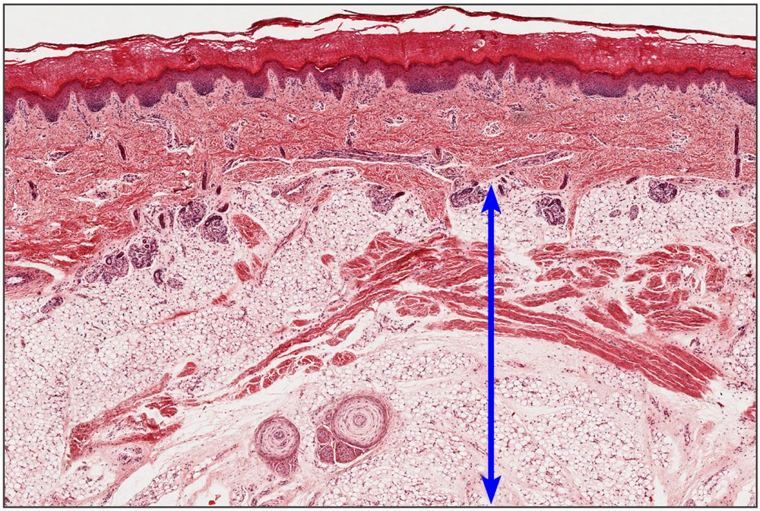
The hypodermis (subcutis, hipodermis) is:
- made of loose connective tissue;
- rich in adipose cells (adipocytes), grouped into lobules;
- extensively vascularized;
- the home of Pacinian corpuscles, sensory endings that detect pressure.
It insulates the body, stores energy, cushions against blows and lets the skin slide over the structures beneath it. Strictly speaking it is not part of the skin (it corresponds to the superficial fascia of anatomy), but it is studied with it. Its thickness varies greatly between sites and people; the layer of fat in it is called the panniculus adiposus.

Sensory nerve endings

The skin's nerve endings are either free (bare branches of the axon) or attached to a special structure: a Merkel cell, or a capsule of connective tissue and flattened cells. The capsule shapes what the ending responds to.
Corpusculum Merkeli (Merkel's disc)
- Unencapsulated.
- Free nerve endings attached to Merkel cells.
- Located in the basal layer of the epidermis.
- A receptor for light touch / tactile sensation, particularly sustained touch and fine texture (numerous on the fingertips).
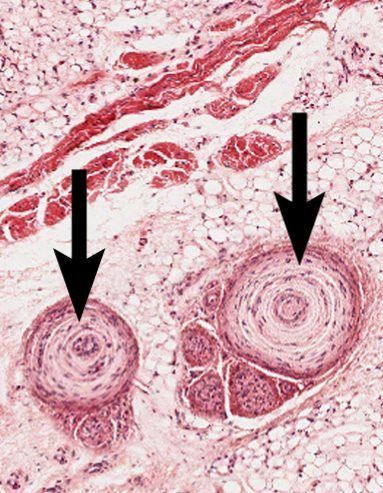
Corpusculum Vater-Pacini (Pacinian corpuscle)

- Oval, onion-shaped.
- A nerve ending surrounded by a capsule of concentric layers (lamellae), made of collagen fibers and flattened, endothelial-like cells, with fluid between them.
- Located in the deep dermis and the subcutaneous tissue.
- A receptor for crude touch, sustained pressure and vibration.
- Large (up to 1–2 mm), so it is easy to spot even at low power.
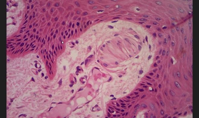
Corpusculum Meissner (Meissner's corpuscle)

- Cylindrical (elongated, oval).
- Branched, encapsulated nerve endings.
- A thin capsule of connective tissue, with flattened cells arranged transversely inside it (like a stack of coins).
- Located in the dermal papillae, just beneath the epidermis.
- A receptor for light touch; abundant on the lips, fingertips, palms and soles.
Corpusculum Ruffini (Ruffini ending)

- Cylindrical, and smaller than Meissner's corpuscles.
- Nerve endings that branch in a spray-like pattern, encapsulated.
- A thin capsule of connective tissue.
- Located in the dermis.
- A receptor for pressure (and for stretch of the skin, which signals joint and finger position).
Corpusculum Krause (Krause end bulb)

- Round / spherical.
- Branched, encapsulated nerve endings.
- A thick capsule of connective tissue.
- Located in the dermis (also in mucous membranes such as the lips and conjunctiva).
- The practicum lists it as a receptor for pressure. Older textbooks called it a cold receptor; its exact role is still debated, so give the practicum's answer in the practicum.
Free nerve endings
The simplest endings: bare branches of sensory axons reaching into the epidermis. They detect pain, temperature and itch, and they are by far the most numerous. A related type wraps around the base of each hair follicle (the root hair plexus), sensing hair movement.
Summary: the five named endings
| Ending | Shape | Capsule | Location | Senses |
|---|---|---|---|---|
| Merkel | Disc on a Merkel cell | None | Basal epidermis | Light touch |
| Meissner | Cylindrical | Thin; transverse flattened cells | Dermal papillae | Light touch |
| Vater-Pacini | Oval, onion-like | Concentric lamellae | Deep dermis, hypodermis | Crude touch, pressure, vibration |
| Ruffini | Cylindrical, small | Thin; spray-like branches | Dermis | Pressure, stretch |
| Krause | Round | Thick | Dermis | Pressure (practicum) |
Two contrasts are asked most often:
- Meissner vs Pacini: Meissner is small, cylindrical and high up in the papillae (light touch); Pacini is large, onion-like and deep (pressure and vibration).
- Meissner vs Ruffini vs Krause: all encapsulated and branched. Meissner is in the papillae; Ruffini is small with a spray of branches; Krause is round with a thick capsule.