Chapter 5: Epithelial Junctions, Surface Specializations & Glands
Chapter 1 covered the two classification axes tested most often — layer count and surface cell shape. This chapter covers the material that turns "I can name the epithelium" into "I understand why it looks and behaves that way": how epithelial cells stick together, what their surfaces are built for, and how epithelium folds itself into glands.
General features, in one place
- Epithelial cells are polyhedral and packed tightly together with very little extracellular matrix between them.
- That tight packing is held together by adhesion molecules, membrane interdigitation, and a junctional complex (below) — not just proximity.
- Every epithelial cell sits on a basement membrane, which separates the epithelium from the connective tissue underneath.
- Epithelium forms continuous sheets covering external and internal body surfaces, and also forms the secretory units of exocrine glands.
- It's a selective barrier, and it is avascular — it receives its nutrients by diffusion from the connective tissue below, not from vessels of its own.
Three classification criteria
Chapter 1 introduced layers and surface shape. There's a third axis:
- Number of cell layers — simple (one layer) or stratified/complex (two or more).
- Surface cell shape — squamous, cuboidal, or columnar.
- Surface specialization — plain, ciliated, or keratinized/cornified.
A full identification — "stratified squamous, keratinized" or "simple columnar, ciliated" — uses all three.
Apical, lateral, and basal surfaces
Every epithelial cell has three functionally distinct faces:
- Apical surface — faces the lumen or external surface. This is where microvilli, cilia, and stereocilia project from.
- Lateral surface — faces the neighboring cell. This is where the junctional complex sits.
- Basal surface — faces the basement membrane, anchored by hemidesmosomes.
The junctional complex
- Tight junction (zonula occludens) — seals the space between cells, controlling what can pass between them (the paracellular route).
- Zonula adherens — a belt-like junction just below the tight junction, anchoring the actin cytoskeleton of neighboring cells.
- Desmosome (macula adherens) — a spot-like "rivet" that anchors intermediate (keratin) filaments between cells, providing mechanical strength (see also Chapter 1's flashcards on this).
- Gap junction — channels called connexons, each built from six transmembrane connexin proteins, bridge the 2–4 nm gap between cells. Small molecules and inorganic ions (up to ~1.5 nm) pass directly from one cell's cytoplasm to the next; larger molecules and negatively charged ions cannot.
- Hemidesmosome — found only on the basal surface, anchoring the epithelial cell to the basement membrane (not to another cell).
Surface specializations
- Microvilli — finger-like projections of the apical plasma membrane, 0.5–1.0 µm long, specialized for absorption. They increase apical surface area roughly 30-fold — the defining feature of absorptive epithelium like the small intestine's brush border.
- Stereocilia — unusually long, immobile microvilli, easily seen with a light microscope. Found in small numbers in specific parts of the male reproductive tract, classically the epididymis, where they assist absorption.
- Cilia — motile projections of the apical surface, found on epithelium of the respiratory tract and the female reproductive tract (fallopian tube). A single cell can carry up to 300 cilia, each up to 10 µm long (about half the cell's height). Cilia beat in a synchronized, wave-like rhythm that drives fluid or mucus in one consistent direction — moving mucus toward the throat in the airway, or moving the ovum from ovary toward uterus in the fallopian tube.
Goblet cells
Goblet cells are modified columnar cells scattered among the columnar cells of the respiratory and GI tract. They synthesize and secrete mucin, which hydrates into protective mucus (see Chapter 1's flashcards for more).
Glandular epithelium
Epithelium doesn't just cover surfaces — it also folds inward during development to form glands.
- Exocrine glands keep a duct connecting them to an epithelial surface.
- Endocrine glands lose that duct during development and secrete hormones directly into surrounding capillaries (e.g. the thyroid).
Exocrine glands: classified by structure
Structurally, exocrine glands are classified by their duct (simple = unbranched, compound = branched) and by the shape of the secretory unit (tubular, alveolar/acinar, or a tubuloalveolar mix). Chapter 1 already covers the complementary classification by secretion mechanism — merocrine, apocrine, and holocrine.
Distribution — where each epithelium type is found
| Type | Subtype | Classic location |
|---|---|---|
| Squamous | Simple | Blood vessels (endothelium), body cavities (mesothelium), alveoli, Bowman's capsule, loop of Henle |
| Squamous | Stratified, non-keratinized | Oral cavity, esophagus, anus, cervix, vagina, cornea |
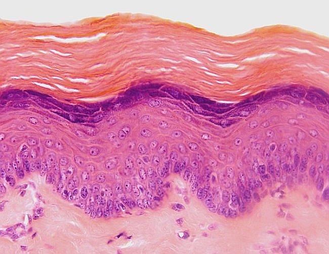
| Squamous | Stratified, keratinized | Epidermis (skin) |
| Cuboidal | Simple | Kidney collecting tubules, thyroid follicles, small exocrine duct, ovary surface |
| Cuboidal | Stratified | Larger ducts of exocrine glands (e.g. sweat and salivary gland ducts) |
| Columnar | Simple | Gall bladder, kidney collecting ducts, endocervix, intestine |
| Columnar | Pseudostratified, ciliated | Respiratory tract (nose, trachea, bronchi) |
| Columnar | Simple, ciliated | Fallopian tubes |
| Columnar | Stratified | Large excretory ducts, male urethra (pars cavernosa) |
| Transitional | — | Lower urinary tract — renal pelvis, ureters, bladder, urethra |
Microscopic atlas
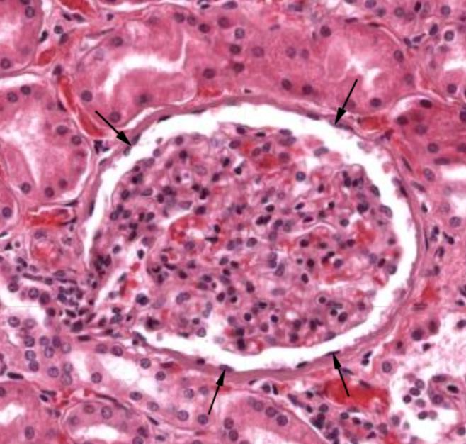
Simple squamous — parietal layer of Bowman's capsule, renal corpuscle (arrows mark the thin squamous cells).
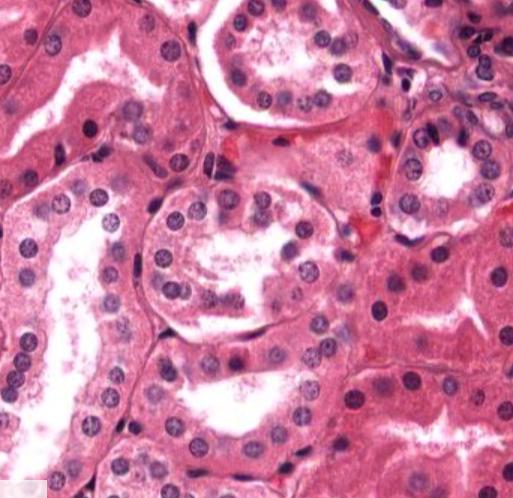
Simple cuboidal — renal tubules.
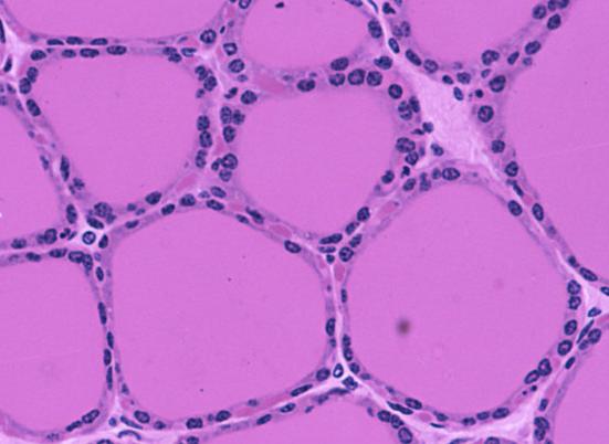
Simple cuboidal — follicular cells of the thyroid gland.
Stratified squamous, non-keratinized — esophagus.
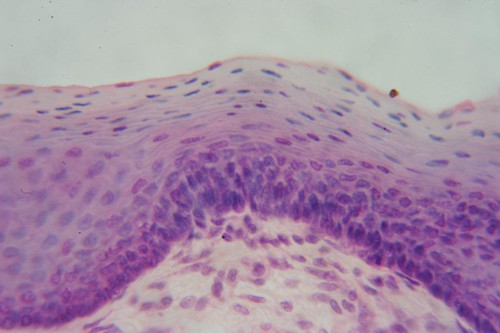
Stratified squamous, keratinized — skin (epidermis), with its dense pink keratin layer on top.
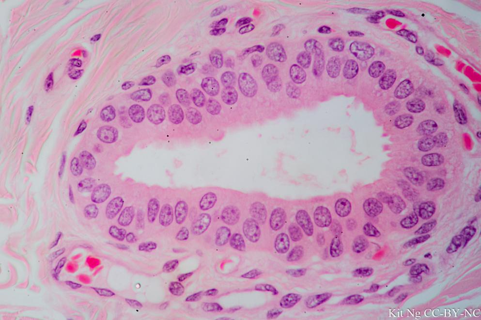
Stratified cuboidal — excretory duct of a salivary gland. (Micrograph: Kit Ng, CC BY-NC.)
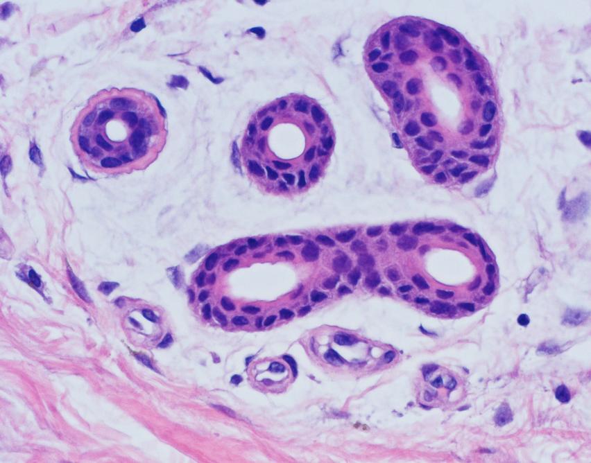
Stratified cuboidal — excretory ducts of sweat glands.
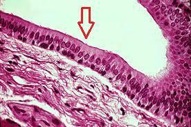
Stratified columnar — male urethra, pars cavernosa.
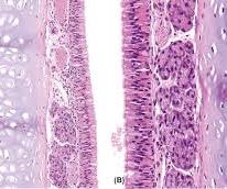
Pseudostratified ciliated columnar — tracheal mucosa.
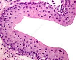
Transitional epithelium — bladder, relaxed (dome-shaped surface cells).
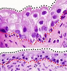
Transitional epithelium — bladder, relaxed vs. distended (surface cells flatten as the wall stretches).
Key teaching point
Every fact in this chapter answers the same underlying question: why does this epithelium look the way it does, here? Junctions explain how a sheet stays a sheet. Surface specializations explain what a surface is for — absorption (microvilli), movement (cilia), or protection (keratin). And gland structure explains what happens when a sheet of epithelium folds inward instead of staying flat. Read the distribution table as a set of worked examples of all three ideas at once.