Chapter 3: Skin Structure & the Epidermis
The skin (integument) is the body's largest organ: about 15% of body weight and close to 2 m² in an adult. It keeps water in and microbes out, blocks ultraviolet light, helps control body temperature, makes vitamin D, and is packed with sensory endings. The integumentary system is the skin plus its appendages: hair, nails and glands (Chapter 5).
This chapter covers the overall layout of the skin and its outer layer, the epidermis: its strata, the difference between thick and thin skin, and the four kinds of cells that live in it besides keratinocytes.
Layers of the skin

The skin has two layers, and it sits on a third:
| Layer | Tissue | Origin |
|---|---|---|
| Epidermis | Keratinized stratified squamous epithelium | Surface ectoderm |
| Dermis | Connective tissue: a thin loose papillary layer over a thick dense reticular layer | Mesoderm |
| Hypodermis (subcutis) | Loose connective tissue rich in fat | Mesoderm; not strictly part of the skin |
The boundary between epidermis and dermis is not flat. Epidermal ridges (rete ridges) project down into the dermis and interlock with dermal papillae projecting up, like the teeth of a zipper. This increases the area of contact, anchors the epidermis against shearing forces, and brings capillaries close to the epidermis, which has no blood vessels of its own: it is nourished by diffusion from the dermis. On the fingertips the ridges form the fingerprint pattern.

The epidermis
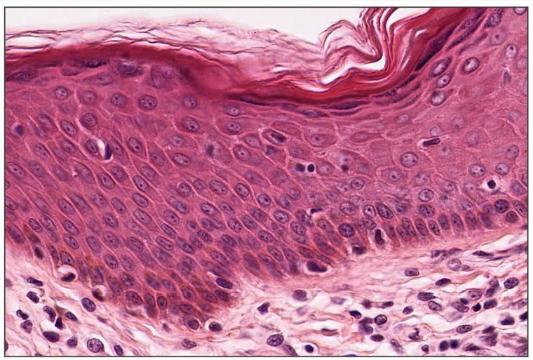
The epidermis is keratinized stratified squamous epithelium. Its main cell is the keratinocyte, which is born in the deepest layer and pushed toward the surface, changing as it goes: it fills with keratin, loses its nucleus and organelles, and dies as a flat, tough scale that is finally shed. That whole journey (keratinization) takes about two to four weeks.
The strata, deep to superficial
1. Stratum basale (stratum germinativum)
- A single layer of cuboidal or columnar, basophilic cells sitting on the basal lamina, to which they are attached by hemidesmosomes.
- Joined to each other on their apical and lateral surfaces by desmosomes.
- High mitotic activity: it contains the progenitor (stem) cells that give rise to all keratinocytes.
- Melanocytes and Merkel cells sit among the basal cells.
2. Stratum spinosum
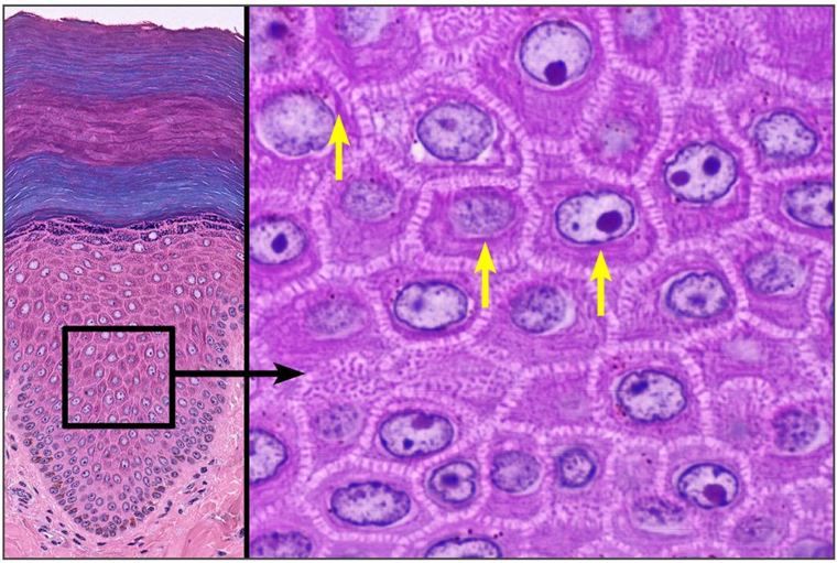
- Several layers of polygonal cells with central nuclei.
- In routine sections the cells shrink apart but stay joined at their desmosomes, so each cell appears covered with short "spines", the intercellular bridges that give the layer its name.
- Bundles of keratin intermediate filaments, tonofibrils, run into the desmosomes and are visible as fine cytoplasmic strands.
- Langerhans cells are found mainly here.
- Together, the basale and spinosum are called the stratum Malpighii, the living, dividing part of the epidermis.

3. Stratum granulosum
- Three to five layers of flattened cells full of dark, basophilic keratohyalin granules.
- The cells also secrete lamellar granules of lipid into the spaces between cells, which make the skin waterproof.
- Above this layer the cells die: nuclei and organelles break down.
4. Stratum lucidum
- Several layers of flattened, pale cells.
- Anucleate (no nuclei).
- The cytoplasm contains eleidin, a transformation product of keratohyalin, which makes the layer look clear and glassy (lucidum, "bright").
- Found only in thick skin.
5. Stratum corneum
- 15–20 layers of flattened, dead, anucleate cells (corneocytes).
- The cytoplasm is filled with refractile filamentous keratin.
- The superficial cells are continually shed (desquamation) as the last step of keratinization.
Thick skin and thin skin

"Thick" and "thin" describe the epidermis, not the whole skin.
| Thin skin | Thick skin | |
|---|---|---|
| Where | Most of the body | Palms and soles |
| Stratum corneum | Thin | Very thick |
| Stratum lucidum | Absent | Present |
| Hair and sebaceous glands | Present | Absent |
| Sweat glands | Present | Very numerous |
| Epidermal ridges | Low | Deep (fingerprints) |
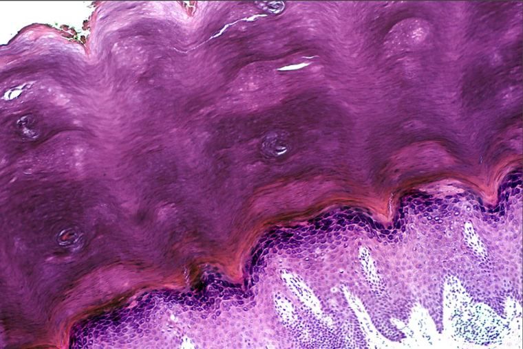
The quick rule at the microscope: a stratum lucidum or a stratum corneum thicker than the rest of the epidermis means thick skin; hair follicles in the section mean thin skin.
Cells of the epidermis

Keratinocytes make up about 90% of the epidermis. The others:
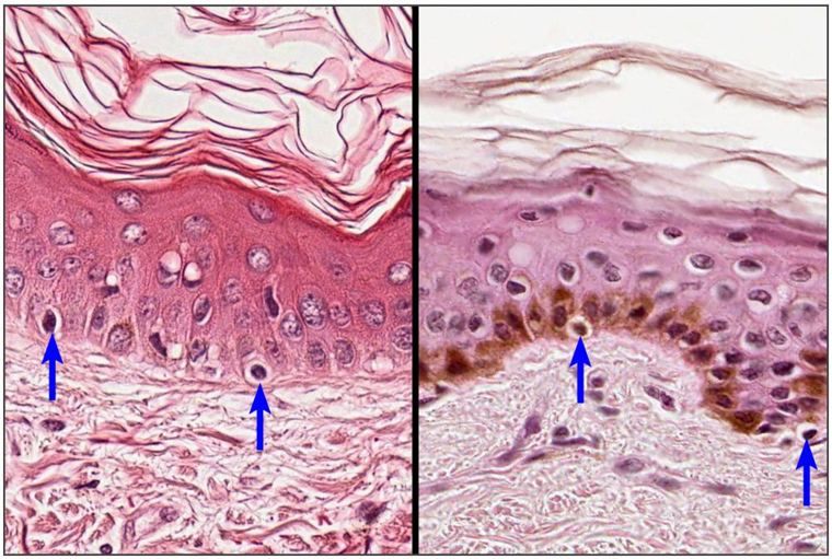
Melanocytes
- A round cell body with long cytoplasmic processes (dendrites).
- Located between the cells of the stratum basale.
- They produce the pigment eumelanin in organelles called melanosomes and pass it through their processes to the surrounding keratinocytes, where it forms a cap over the nucleus that shields the DNA from ultraviolet light.
- Skin color depends on how much melanin is made and transferred, not on the number of melanocytes, which is similar in light and dark skin.
- In H&E they appear as pale "clear" cells in the basal layer.

Langerhans cells
- Morphologically similar to melanocytes, with long dendritic processes, but they do not produce melanin.
- Found mainly in the stratum spinosum.
- Immune cells: they act like macrophages, capturing antigens that cross the skin and carrying them to lymph nodes to present to T cells.
- In H&E they are hard to see; special stains (gold chloride, or immunostaining for CD1a) show them as dark cells with branching processes.
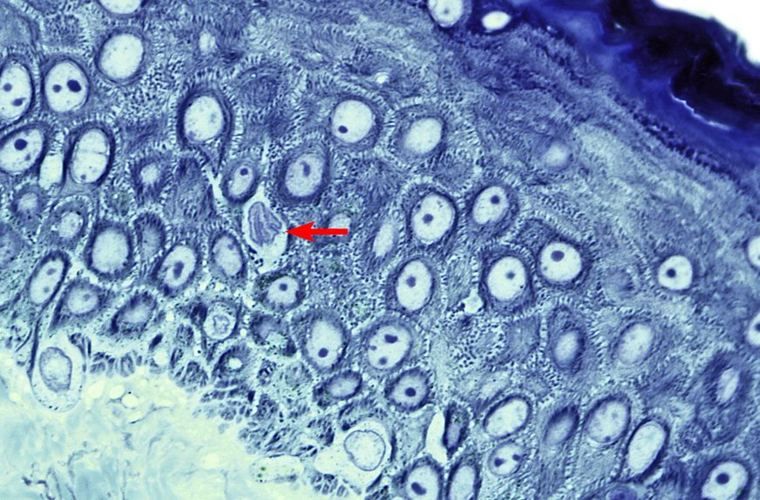
Merkel cells
- Morphologically similar to keratinocytes, but their cytoplasm contains small, dense granules (neurosecretory granules).
- Found in the basal layer, especially in thick skin (fingertips).
- A free nerve ending expands into a disc at the base of each cell; together they form the Merkel corpuscle (Merkel's disc), a mechanoreceptor for light, sustained touch (Chapter 4).
Lymphocytes
- Round cells with a round nucleus of coarse, densely packed chromatin.
- Most are immature T lymphocytes, whose maturation in the epidermis is supported by keratinocytes.
- With Langerhans cells and keratinocytes, they form the skin's immune system, SALT (skin-associated lymphoid tissue).
| Cell | Where | What it does | How to recognize it |
|---|---|---|---|
| Keratinocyte | All layers | Keratin, barrier | The ordinary cells of every stratum |
| Melanocyte | Stratum basale | Eumelanin | Pale cell among basal cells, dendrites |
| Langerhans cell | Stratum spinosum | Antigen presentation | Dendritic, no melanin; special stains |
| Merkel cell | Stratum basale | Touch receptor (with its nerve ending) | Dense granules; thick skin |
| Lymphocyte | Epidermis | Immunity (SALT) | Small round cell, dense nucleus |